
Introduction
T lymphocytes (T cells) play a specialized, critical role in the antigen-specific immune response. When activated, these cells can recognize and directly kill virally infected or malignant cells. Although T cells can be isolated efficiently from blood and grown ex vivo for cancer therapy (e.g., in adoptive cell therapy with chimeric antigen receptors), they are difficult to manipulate genetically and to efficiently expand to therapeutically relevant numbers.
In vitro T-cell expansion can be boosted by the addition of growth factor interleukin 2 (IL-2) in combination with anti-CD3 monoclonal antibody (OKT3 clone). CD3 pathway activation renders T cells susceptible to IL-2 receptor stimulation, triggering proliferation. Introducing anti-CD3 antibodies disrupts antigen receptors on the surface of T cells that are associated with the CD3 complex, inducing antigen receptor activation (Tsoukas et al. 1985). In conjunction with IL-2 and anti-CD3 antibody, RetroNectin reagent has been shown to further increase the expansion of naïve CD8+ T cells from peripheral blood mononuclear cells (PBMCs), and to improve the expansion and post-transplant persistence of genetically modified T cells (Yu et al. 2008). RetroNectin improves CD8+ T-cell expansion through the stimulation of the integrins VLA-4 and VLA-5, enhancing effector multifunctionality and in vivo memory formation (Hosoi et al. 2014).
RetroNectin Recombinant Human Fibronectin Fragment and RetroNectin GMP grade have widespread use as enhancers of retroviral or lentiviral gene transfer into hard-to-transduce cell types. Here, we present data on an additional application, the improved expansion of T cells from PBMCs and increased production of naïve T cells, using a combination of our products: RetroNectin reagent, CultiLife culture bags, anti-CD3 antibody, and T-cell culture medium.
Results
PBMCs were isolated from the peripheral blood of two healthy donors, cultured with anti-CD3 antibody, and assayed for overall T-cell expansion and for the proportion of naïve T cells, as defined by the expression of CD45RA and CCR7 (see Methods). The results showed a marked increase in expansion capacity and a higher proportion of naïve T cells with the use of RetroNectin reagent.
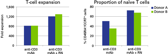
Figure 1. RetroNectin reagent (RN) increases expansion and naïve T-cell fraction of PBMCs stimulated with anti-CD3 antibody and IL-2. Peripheral blood T cells stimulated and expanded in vitro with anti-CD3 antibody (OKT3 clone) and IL-2 for 10 days contained approximately two-fold higher naïve T cells (CD45RA+/CCR7+ phenotype) in the presence of immobilized RN than in the absence of RN.
Conclusions
Anti-CD3/IL-2 activation in combination with RetroNectin reagent dramatically improves T-cell expansion in vitro. This combination results in a higher proportion of naïve T cells, providing high-quality starting material for further research and therapies based on T cells, such as adoptive cell therapy. Read more about ex vivo gene therapy and the use of RetroNectin in clinical settings.
Methods
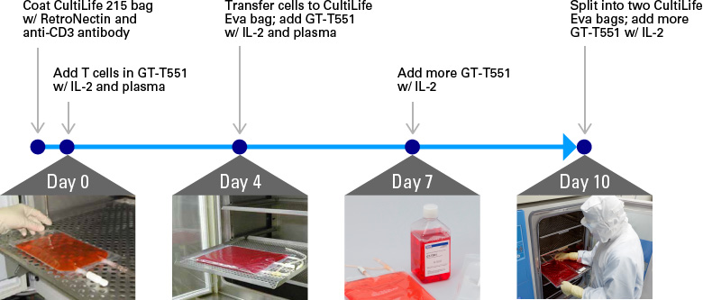
Figure 2. T-cell expansion protocol using RetroNectin reagent, GT-T551 T-cell medium, CultiLife bags, and anti-CD3 mAb.
PBMCs and autologous plasma were prepared using 50 ml of blood from two healthy volunteers with informed consent. PBMCs from each donor were split into two samples to test anti-CD3-antibody- and IL-2-induced expansion with or without RetroNectin reagent (RN). CultiLife 215 bags (CultiLife 215 Culture Bag) were coated with either anti-CD3 antibody plus RN or anti-CD3 antibody alone in PBS for 2 hr, then rinsed three times with PBS. On Day 0, PBMCs were resuspended in 50 ml of GT-T551 medium supplemented with heat-inactivated plasma (0.6% final volume) and added to the coated CultiLife 215 bags. Additional GT-T551 medium supplemented with IL-2 and plasma was added to each CultiLife 215 bag to reach a final volume of 250 ml. The PBMCs were transferred to CultiLife Eva bags (GT-T610 [CultiLife Eva] Culture Bag) on Day 4 and fresh GT-T551 medium (with IL-2 and 0.6% inactivated plasma) was added to bring the volume to 500 ml per bag. An additional 500 ml of medium supplemented with only IL-2 was added on Day 7. On Day 10, cells were split into two CultiLife Eva bags, with fresh medium and IL-2 added to bring the final volume of each bag to 1,000 ml. The cells were harvested on Day 14, the total number of T cells was counted, and the proportion of naïve (CD45RA+/CCR7+) T cells was determined by flow cytometry analysis.
References
Hosoi, H. et al. Stimulation through very late antigen-4 and -5 improves the multifunctionality and memory formation of CD8+ T cells. Eur. J. Immunol. 44, 1747–58 (2014).
Tsoukas, C. D. et al. Activation of resting T lymphocytes by anti-CD3 (T3) antibodies in the absence of monocytes. J. Immunol. 135, 1719–23 (1985).
Yu, S. S. et al. In vivo persistence of genetically modified T cells generated ex vivo using the fibronectin CH296 stimulation method. Cancer Gene Ther. 15, 508–16 (2008).